Did CHIDA et al. show us the catastrophic risk of rupture of Vertebral Artery Dissecting Aneurysm after mRNA technology based COVID gene injection vaccine (Moderna & Pfizer), looking at 2 cases? Yes!
SOURCE:
https://www.ncbi.nlm.nih.gov/pmc/articles/PMC9119691/
Researchers reported ‘two cases of ruptured vertebral artery dissecting aneurysm (VADA) immediately after mRNA (mRNA) technology based CIVID gene injection.
Case 1:
a 60-year-old woman experienced sudden headache 3 weeks before her first dose of the Moderna mRNA-1273 COVID-19 vaccine. Magnetic resonance imaging showed dilatation of the right vertebral artery (VA) without intracranial hemorrhage. A day after the vaccination, she developed subarachnoid hemorrhage with pulmonary effusion due to a ruptured right VADA. She underwent endovascular internal trapping and parent artery occlusion under general anesthesia.
Case 2:
a 72-year-old woman with a previous history of the left VA occlusion due to arterial dissection developed subarachnoid hemorrhage 7 days after the first dose of the Pfizer-BioNTech BNT162b2 COVID-19 mRNA vaccine due to a ruptured right VADA and underwent stent-assisted coil embolization under general anesthesia.’
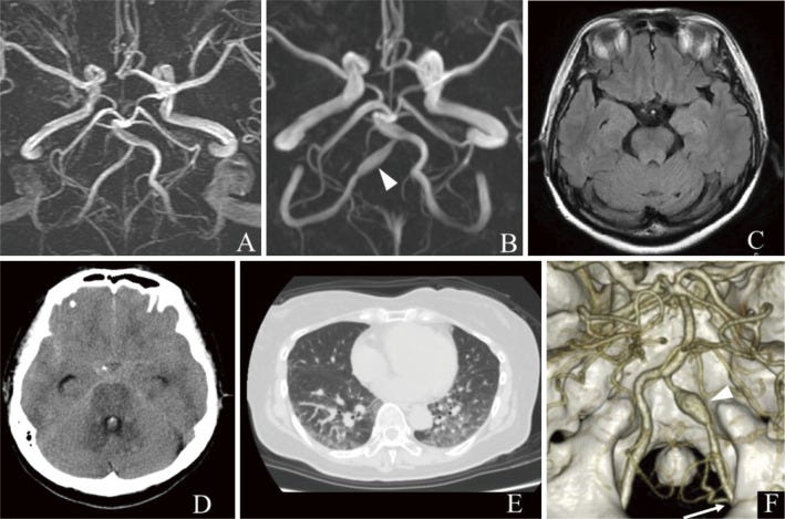
(A) Magnetic resonance angiography (MRA) performed 10 years previously shows normal right vertebral artery (VA). (B) MRA performed 3 weeks before the vaccination shows dilatation of the right VA (arrowhead). (C) Fluid-attenuated inversion recovery magnetic resonance imaging (MRI) performed on the same day shows no intracranial hemorrhage. (D) Head computed tomography (CT) performed 1 day after the vaccination shows diffuse subarachnoid hemorrhage. (E) Chest CT performed on the same day shows bilateral pulmonary effusion. (F) Three-dimensional CT angiography (3DCTA) performed on the same day shows dissecting aneurysm in the right VA (arrowhead). The right posterior inferior cerebellar artery (PICA) originated from the extradural segment of the VA (arrow).
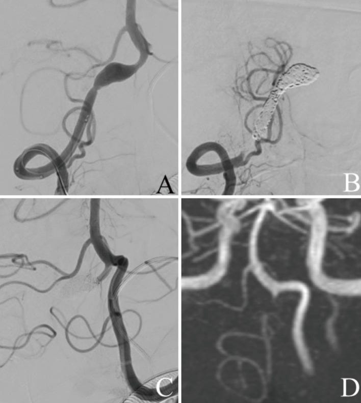
(A) Frontal view of the right vertebral artery angiography (VAG) before internal trapping shows dissecting aneurysm in the right VA. (B) Frontal view of the right VAG after internal trapping reveals that the dissecting lesion was occluded just distal to the origin of the right posterior inferior cerebellar artery (PICA). (C) Frontal view of the left VAG after internal trapping also reveals that the dissecting lesion was occluded. (D) Postoperative magnetic resonance angiography (MRA) performed 2 weeks after treatment reveals that the dissecting lesion of the right VA was not visualized.
(A) Head computed tomography (CT) performed 7 days after vaccination shows massive subarachnoid hemorrhage in the posterior cranial fossa. (B) Three-dimensional CT angiography (3DCTA) performed on the same day shows a dissecting aneurysm with bleb-like protrusion on the right vertebral artery (VA). (C) Magnetic resonance angiography (MRA) performed 1 year previously shows caliber change of the right VA, retrospectively, suggesting chronic arterial dissection.
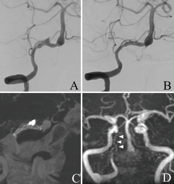
(A) Frontal view of the right vertebral artery angiography (VAG) before stent-assisted coil embolization shows dissecting aneurysm in the right vertebral artery (VA) involving the right posterior inferior cerebellar artery (PICA). (B) Frontal view of the right VAG after stent-assisted coil embolization shows coil occlusion of the aneurysmal sac and patency of the right VA and right PICA. (C) Cone beam computed tomography (CT) image shows the bladed stent placed across the dissecting lesion and coils deployed into the sac. (D) Postoperative magnetic resonance angiography (MRA) performed 2 weeks after treatment shows that the dissecting aneurysm is not visualized and the right PICA is patent.


I just have to say it. We know these jabs maim and kill already! We don't need further research studies!
We need JUSTICE!