LNPs, exosomes, extracellular vehicles: did evidence show early on that lipid nano particles (LNPs) exchange more easily in small diameter vessels with low flow rate i.e., capillaries & small vessels?
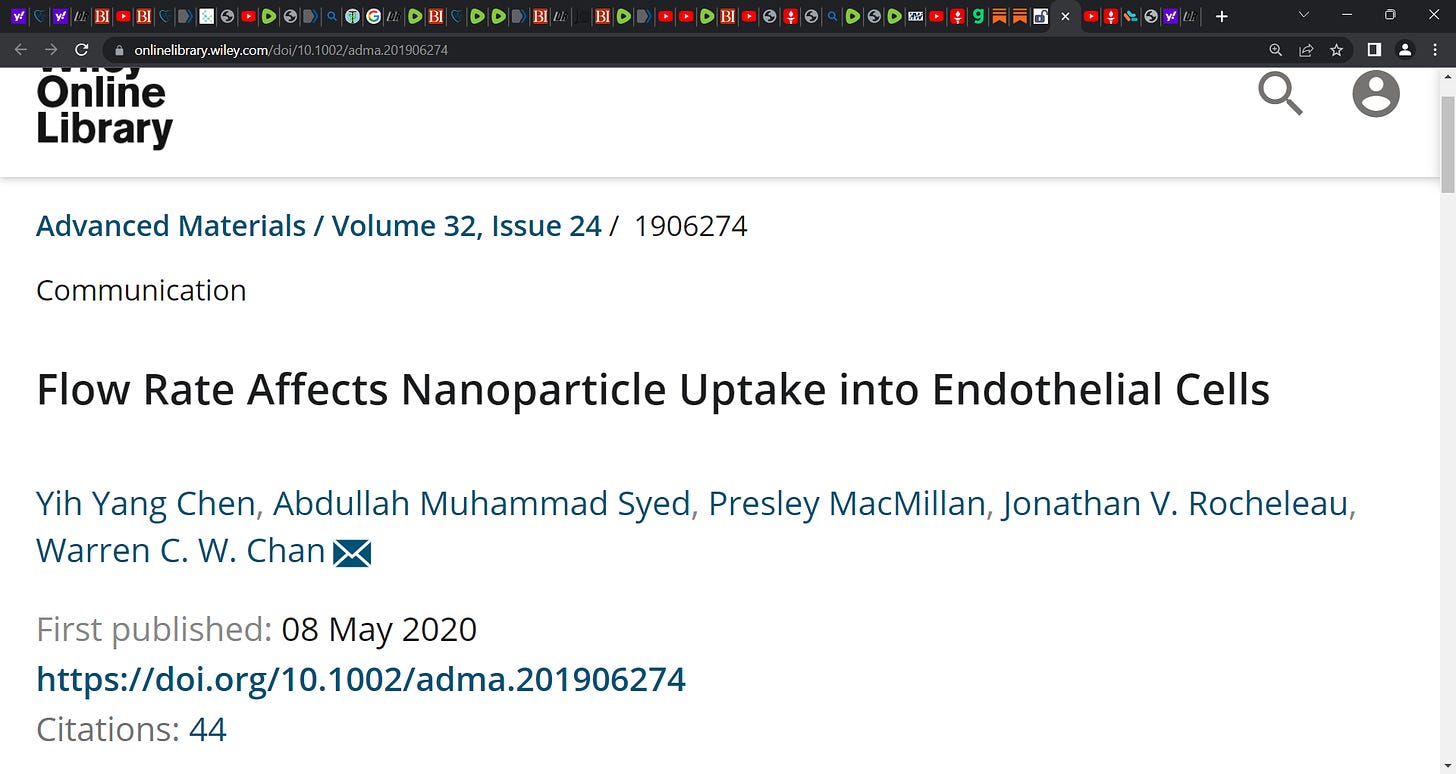
Yes! Chen et al. showed us this via how flow Rate Affects Nanoparticle Uptake into Endothelial Cells
https://onlinelibrary.wiley.com/doi/10.1002/adma.201906274
‘Injected nanoparticles travel within the blood and experience a wide range of flow velocities that induce varying shear rates to the blood vessels.
Endothelial cells line these vessels, and have been shown to uptake nanoparticles during circulation, but it is difficult to characterize the flow-dependence of this interaction in vivo.
Here, a microfluidic system is developed to control the flow rates of nanoparticles as they interact with endothelial cells. Gold nanoparticle uptake into endothelial cells is quantified at varying flow rates, and it is found that increased flow rates lead to decreased nanoparticle uptake.
Endothelial cells respond to increased flow shear with decreased ability to uptake the nanoparticles. If cells are sheared the same way, nanoparticle uptake decreases as their flow velocity increases. Modifying nanoparticle surfaces with endothelial-cell-binding ligands partially restores uptake to nonflow levels, suggesting that functionalizing nanoparticles to bind to endothelial cells enables nanoparticles to resist flow effects.’


Vaccine manufacturers knew of the harm lipid Nanoparticles caused in the late 80s-early 90s. Because they tried them back then, then dropped the technology like a hot potato when animals & people got hurt.
Those at risk for decreased microvascular vasomotion are lightning rods for the LNP (and spike protein) damage to endothelium due to baseline stagnation of microvascular blood flow and diminished vasomotion:
Evaluation of the microcirculation in vascular disease
Christopher J. Abularrage, MD,a Anton N. Sidawy, MD,a Gilbert Aidinian, MD,b Niten Singh, MD,a Jonathan M. Weiswasser, MD,a and Subodh Arora, MD,c Washington, DC
Insufficient blood flow through end-resistance arteries leads to symptoms associated with peripheral vascular disease. This may be caused in part by poor macrocirculatory inflow or impaired microcirculatory function. Dysfunction of the microcirculation occurs in a similar fashion in multiple tissue beds long before the onset of atherosclerotic symptoms. Impaired microcirculatory vasodilatation has been shown to occur in certain disease states including peripheral vascular disease, diabetes mellitus, hypercholesterolemia, hypertension, chronic renal failure, abdominal aortic aneurysmal disease, and venous insufficiency, as well as in menopause, advanced age, and obesity. Microcirculatory structure and function can be evaluated with transcutaneous oxygen, pulp skin flow, iontophoresis, and capillaroscopy. We discuss the importance of the microcirculation, investigative methods for evaluating its function, and clinical applications and review the literature of the microcirculation in these different states. ( J Vasc Surg 2005;42:574-81.)
This is why I must keep sharing what I’ve learned about circulation and the effect BEMER has to stimulate muscle for better flow of blood through the Microcirculation.
An old saying is “a rolling stone gathers no moss”, and perhaps better rolling blood cells will potentially reduce the risk LNPs from gathering in endothelium, IMHO.